Project description and aims
The DOA project aims at studying and conserving the human remains and the painted cartonnage case of Ta-Sherit-en-Imen (Amun’s little one).
Project short description
The mummy dates to the Third Intermediate Period (1069-664 BC) and was brought to the shores of Lago Majore by the Swiss collector Zaccaria Zanoli at the end of the 19th century. It was exhibited together with the Zanoli collection at the Palazzo Communale of Brissago from 1916 to the 1960s. After more than a hundred years of non-controlled exhibition and storage, the mummy and its cartonnage case are in dire need of conservation.
The complete study of the human remains will not only help to identify the age and medical history of Ta-Sherit-en-Imen, but will also give valuable information on the embalming techniques that were used.
In order to devise adapted conservation measures all materials present (human remains, textiles, cartonnage, polychrome layers) need to be identified and characterized.
Adapted conservation measures will allow the preservation of all materials and bring back dignity to the body that was prepared for eternity approximately 2800 years ago.
At the end of the project, Ta-Sherit-en-Imen will be placed back into her cartonnage case and integrated into the new permanent exhibition of the Kulturama of Zurich.
Timeline and intermediate reports
March 2021 | Temporary stabilization with consolidation of cartonnage case for transport and deposition of the mummy and the two cartonnage case parts
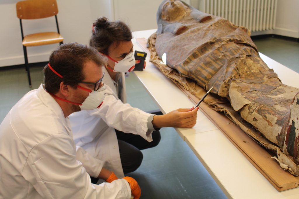
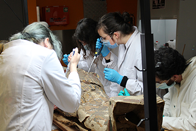
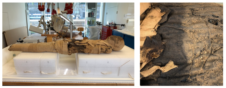
When the cartonnage case was cut open at the beginning of the 20th century its structural integrity was lost. Years of manipulation uncontrolled storage have left a distorted and weakened two-piece cartonnage case with the mummy . The result was delamination, creases and tears in the cartonnage case as well as lifting and loss of paint (Figs. 1 & 2). The extreme fragility of the cartonnage case made handling and transport from Brissago to Neuchâtel impossible.
Not wanting to introduce a consolidant into the paint layers before having the opportunity to study them we looked for a temporary consolidant that could be removed at a later date without leaving any residues. Preliminary analysis of the polychrome layers at the HKB by Dr. S. Zumbühl & Dr. N. Scherrer confirmed the absence of non-polar components. As a result, cyclododecane (C12H24) was chosen as a temporary consolidant. At room temperature cyclododecane, a saturated cyclic alkane, is a white waxy solid with slight odor. Applied in a melt or dissolved in a non-polar solvent it can consolidate fragile surfaces for a finite amount of time. As it sublimates at room temperature, the adhesive will diminish over days and completely disappear over weeks.
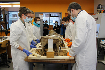
Cyclododecane was applied to the polychomy of the cartonnage with micropipettes in a melt (58°C) with small addition of petroleum ether (100-140°C boilng point) to slow down its solidification (Fig. 3). It was applied before transport and again some weeks later when the upper part of the cartonnage was lifted off the mummy with the help of a custom-made frame (Fig. 4).
March 2021 | Photogrammetry
Photogrammetry is a low-tech technique that can be used to create a precise 3D rendering of an artefact without having to resort to traditional molding techniques. It is of particular interest for very delicate structures that cannot be handled during the process. Objects of all sizes can be treated. The resulting 3D model can be used for the off-location study of the artefact, for establishing condition reports, for monitoring surface alterations, for creating adapted supporting structures for storage and transport as well as the recreation of missing parts.
Equally of interest is the exact rendering of the surface topography that can be viewed with or without colour, allowing a greater precision when assessing surface textures. UV and infra-red images can be integrated in a second step and superposed on the existing surface topography.
For the DOA project the aim was to carefully document the initial condition of the cartonnage case before any intervention was made. This was done in order to document possible changes during transport or handling and to permit a high resolution restitution of the complex surface for an initial condition report. For that 800 high-resolution photographs were taken from various angles by Andreas Hochuli (HKB). These were uploaded to the Metashape (Agisoft) software that reconstructed each photograph, transforming them into scaled models with approximately 120 million surface points. The result is a high-precision virtual model that can be examined from all sides and allows to zoom into areas of particular interest.
The technique will also be used at the end of the project when an adapted supporting structure will have to be made for the upper cartonnage case.
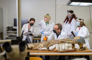
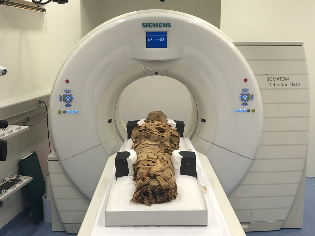
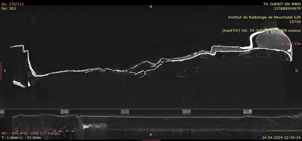
April 2021 | Computed tomography of the mummy
The mummy of Ta-sherit-en-Imen was scanned on April 6 2021 by the team of Dr. Frank Rühli and Patrick Eppenberger of the Swiss Mummy Project, Institute of Evolutionary Medicine of the University of Zurich (IEM). 3,938 sectional axial images with a slice interval of 0.4 mm and a slice thickness of 0.6 mm were reconstructed from the acquired raw data and used to evaluate the findings. The interpretation of the mummification process was completed by Dr. Jonathan Elias from the Akhmim Mummy Studies Consortium (AMSC). A sample of thigh bone was removed, and C-14 dated by Dr. Irka Hajdas from the Laboratory of Ion Beam Physics at the ETH Zurich.
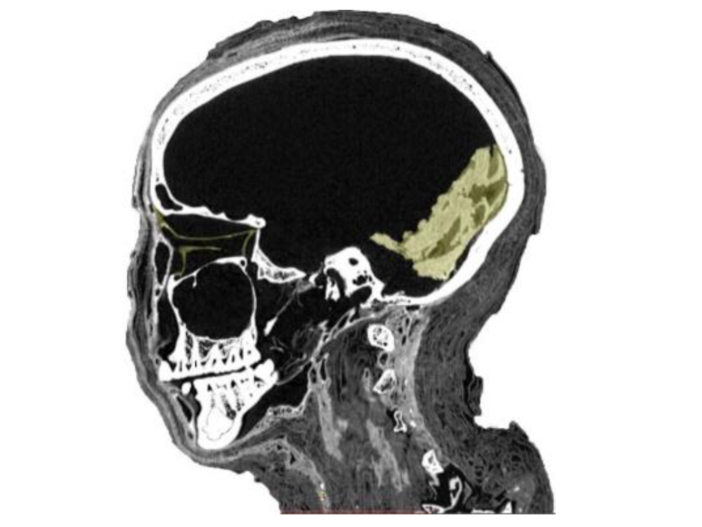
Based on the level of ossification of the cranial sutures, Ta-sherit-en-Imen died as a young adult aged between 27 and 35 years somewhere around 824-780 BC (calibrated C-14 date of bone material). The cause of death was possibly two blows to the frontal-parietal cranium that left two large lesions of the outer compacta, as well as three fracture lines. Since there is no evidence of bone remodeling, she must have died shortly after the incident. The localization of the trauma indicates a foreign impact rather than an accidental lesion.
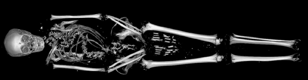
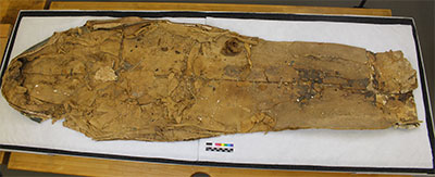
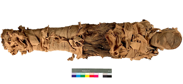
The mummification process was cursory and done without eviscerating internal organs, which is not unusual for the Third Intermediate Period. This led to subsequent disintegration of the soft tissues and the misalignment of the neck and thoracic skeleton (fig. 2). There are no signs for excerebration of the brain and the nasal cells remain intact. The left and right hemispheres of the brain have split at the corpus callosum and fallen on their respective sides (fig. 3).
The individually wrapped arms are arranged angled downwards to the thigh crease with the hands arranged in a relaxed position and nearly touching one another. Three textile packages have been secured to the front of the neck and are held in place by resin coated textile bandages. Such neck packages are frequently found on mummies from that period and seem to be part of the original mummification process. Further textile packages are located on the collapsed left thoracic area as well as in between the legs. Resin was applied onto the bandages twice during the wrapping procedure and can be seen as denser white lines on the scans. The most outer resin coated textile layer can be seen in the middle section of the mummy where outer wrappings were subsequently stripped away. Its hard resinous matter thankfully prevented the complete unwrapping of the mummy.
When Ta-sherit-en-Imen passed away, she was a healthy woman without any visible signs of malnutrition or hard physical work. She was mummified according to the practice of the day, but insufficient dehydration during the mumification process led to subsequent disturbance of some of her skeletal remains.
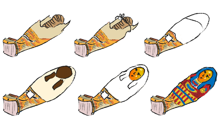
July 2022 | Facial reconstruction
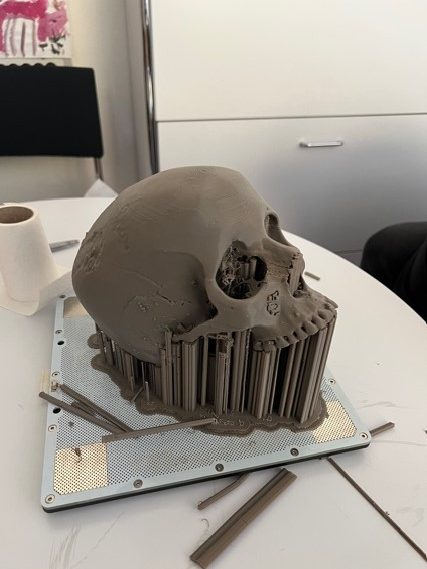
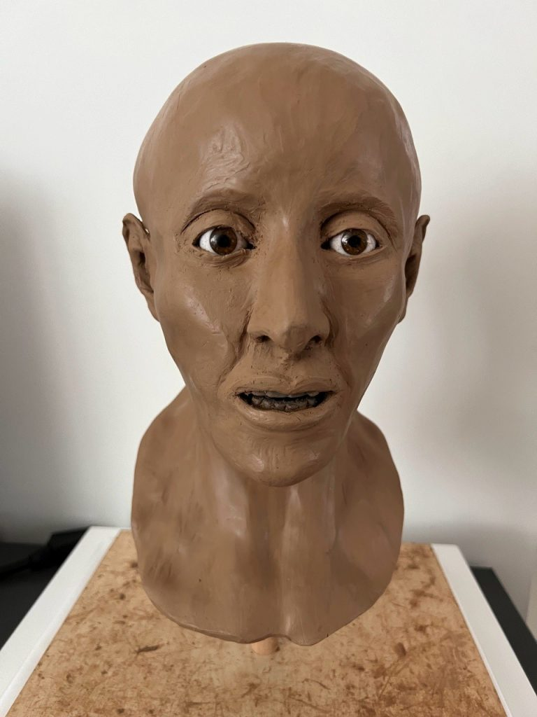
From the computed tomography scan realized at the IEM, a facial reconstruction was elaborated by Dana Ackermann in 2022. The work was done within the framework of a Maturitätsarbeit at the Kantonsschule Limmattal under the supervision Dr. Anna-Katherina Holenweg. The practical reconstruction work was supervised by Dr. med. Patrick Eppenberger at the IEM. If not specified, the images were taken from Achermann’s Maturitätsarbeit and copyright of the Kantonsschule Limmattal.
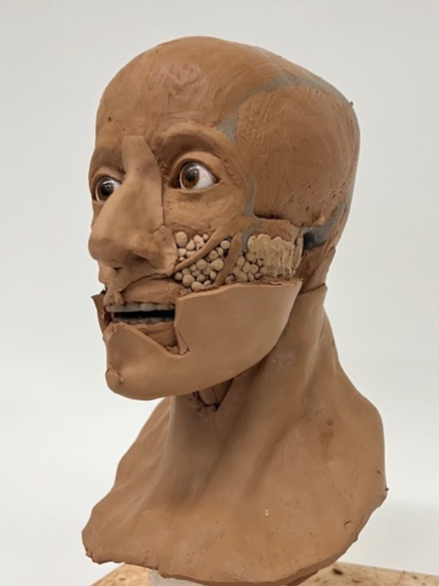
Ackermann used the Gerasimov method (Manchester Method), where muscle and skins are gradually built up by hand over the 3D printed scull. Skin thickness is based on specific data on ethnicity and indicated with tubular spacers that are placed all over the scull. Age, sex, way of life and nutrition are all impact the thickness of muscles, glands and fatty tissues.
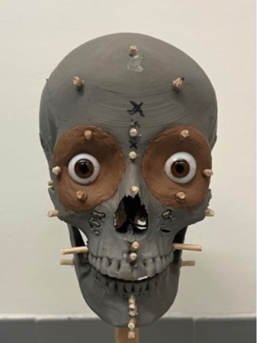
Once the spacers in place, every muscle and gland is carefully modelled by hand. Symmetry, size and form of the nose is reconstructed according to ethnicity, but also on the shape and size of the apertura piriformis, the pear-shaped nasal cavity of the scull. The dental record informs the shape and size of mouth and lips. The ears are the most difficult and least accurate parts, as the scull does not inform us on how they looked like. They are modelled according to available data on ethnicity. The size of the inset glass eyes is chosen according to the aperture of the eye sockets. Once all muscles and glands have been placed, the skin is laid over them in several pieces and joins are smoothed over.
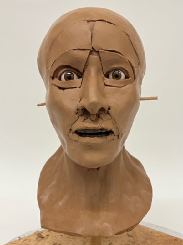
The degree of finish is the one that is usually used in forensic investigations in order to visualize and identify human remains. At this stage modelling marks are still visible; the skin tone is the one of the modelling material, and no attempt is made for the reconstruction of facial hair. This said, it gives us a very real first impression of what Ta-Sherit-en-Imen looked like some 2800 years ago.
The careful and excellent work of Dana Ackermann can only be applauded, more particularly as this was her first attempt of a facial reconstruction.
June 2023 | Conservation of the mummy and its textile bandages
In the late 19th century, the mummy of Ta-Sherit-en-Imen was partially unwrapped, exposing the resin coated bandages of the abdominal area. This was most likely done in the search for amulets or to access the mummified body. On the right hip some of the bandages were removed exposing the parchment-like skin. Fortunately, the inner layers were heavily impregnated with resinous matter and prevented any further desecration of the body.
The restoration of the mummy’s bandages was performed by Laura Flückiger as part of her MA thesis (https://sonar.ch/hesso/documents/321908) under the supervision of conservators Valentin Boissonnas (HE-Arc), Mimi Levèque (Massachusetts, USA), and Agniezka Jucker-Woos (Abegg Stiftung Riggisberg).
The conservation project aimed at characterizing the different linen bandages, understanding their sequence, locating the position of loose bandages, and placing as much of them as possible into their original position. The conservation work was done in a designated conservation lab at the HE-Arc that could not be viewed or accessed by the general public.
The fragility of the mummy prohibited turning the body during treatment. A polyethylene foam support was constructed in a series of sections that allowed to temporarily access any section of the mummy while the rest of the body remained well supported.
All textile bands were first carefully cleaned of dust and particles that had accumulated over the years of uncontrolled storage. The presence of mold on some of the textiles necessitated the use of protective gear and the use of HEPA filters for the low suction hoover. The removal of dust and debris was done with fine brushes and tweezers.
Many bandages presented creases and folds that had formed after their unwrapping. These were reshaped by controlled humidification using humidified blotting paper over a semi-permeable Sympatex layer placed over the textile and sandwiched between two sheets of Melinex.
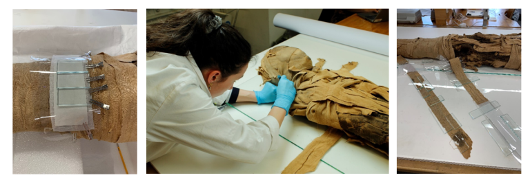
Torn bandages and areas of loss were stabilized by sowing them into two layers of very fine tinted nylon netting. To keep all bandages in place and avoid future damage to fringes and loose threads, a fine tinted nylon gauze was placed over the repositioned bandages. Stitching was done within the nylon mesh. A few loose threads and textile areas that could not be secured mechanically by netting were secured with a 4% methylcellulose in deionized water. Bandages that could not be attributed to a specific position were kept apart as research material.
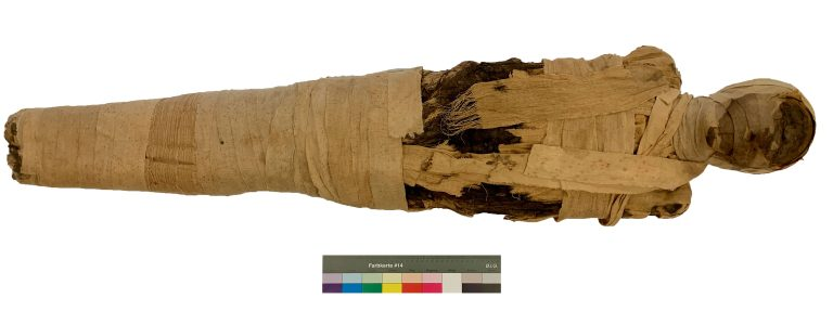
Conserving the mummy of Ta-Sherit-en-Imen was a rare privilege and a can be considered a continuation of the work that embalmers began in ancient Egypt. It gave back dignity to the deceased and enabled her to continue her journey to the beyond and be reborn over and over again. The treatment methodology for Ta-Serit-en-Imen was done with a maximum respect for her person and in accordance today’s conservation ethics. The trauma from the opening of the bandages, at a time when mummies were just another commodity, remains visible and has become an integral part of Ta-Sherit-en-Imen’s history.
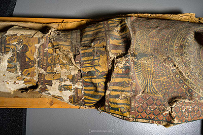
June 2024 | Study of the cartonnage case and its previous restoration
The cartonnage case was cut into two parts at the end of the 19th or early 20th century. This gives us a unique view of its construction method, but also of the previous restoration attempt. The complete study of the cartonnage was the subject of third-year BA dissertation of Marine Roux at the Haute Ecole Arc Conservation-Restauration (https://sonar.ch/global/documents/329807). The visual study was accompanied by a computed tomography scan that was done at the IRIS Radiology Institute in Neuchâtel.
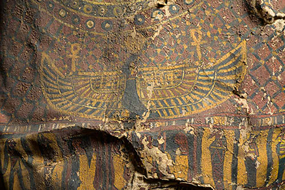
The study revealed how the cartonnage was constructed over a core that was most likely padded with clay and straw to give it a humanoid shape. Layers of linen bandages were wrapped around this core and held in place with an unidentified consolidant. A unique find were blue-and red banded textile strips that had been integrated in a geometric pattern into the core of the cartonnage. Being not visible in the end, they are thought to have had important symbolic value and were an integral part of the mummification process. Similar textile strips have been found on mummies as the last and visible decorative wrapping. The finished cartonnage case was then removed from its core by an incision on its back and covered with a gesso layer and the final paint layers.
The examination also revealed how after excavation the badly damaged upper section of the cartonnage was restored. The remaining original wrappings were reattached with strings and a first cotton textile was adhered over those remains and covered with a gesso layer. Then a Roman-period gilt head from another mummy was positioned on the facial area supported by a block of wood. Below it two wooden planks were attached to represent the two strands of hair. Ancient scrap textiles were used to further wrap and level the reconstructed areas. A final cotton textile was added and painted with the Egyptian motives that can be seen today. In areas original paint layers were also covered by this paint.
We can conclude that this restoration was done to fool potential buyers into believing they were purchasing an original and complete mummy. It is a testament to a flourishing antiquities market that was still going strong in Egypt at the time Zanoli became the owner of the mummy of Ta-Sherit-en-Imen.
Partners and roles
Haute École Arc Conservation-Restauration
The DOA project is coordinated by conservator and lecturer in conservation Valentin Boissonnas. At the HE-Arc CR , MA dissertation topics will be developed around the analysis and conservation of the mummy and its cartonnage case.
Institute of Evolutionary Medicine – University of Zürich
Analysis of the human remains will be performed by the team of Dr. Frank Rühli who is in charge of the Paleopathology and Mummy Studies Group at the Institute of Evolutionary Medicine of the University of Zurich.
Hochschule der Künste Bern – Konservierung-Restaurierung
Conservator and lecturer in conservation Dr. Karolina Soppa will supervise the characterization and conservation the painted cartonnage case and its textiles. Lecturer and conservator Agnieszka Woś Jucker, from the Abegg Stiftung Riggisberg, will be involved with the characterization and conservation of the textile remains
Institute of Evolutionary Medicine – University of Zürich
Analysis of the human remains will be performed by the team of Dr. Frank Rühli who is in charge of the Paleopathology and Mummy Studies Group at the Institute of Evolutionary Medicine of the University of Zurich.
Labor für Ionenstrahlphysik – ETH Zürich
Carbon 14 dating of the different organic remains (human and textile) will be performed by Dr. Irena Hajdas from the Swiss Federal Institute of Technology in Zürich.
Swiss Coffin Project
Study and interpretation of the cartonnage inscriptions will be performed by the Egyptologist Alexandra Küffer. She is co-founder and head of the Swiss Coffin Project.
Ministry of Tourism and Antiquities, Egypt
The DOA project is supported by the Ministry of Antiquities of Egypt. The project includes an exchange of expertise between Egyptian and Swiss conservators and the Haute Ecole Arc will be giving continous education workshops in Cairo.
Press review
Press review: articles, radio program & video
2025
- 13.01.2025 – RSI – Mummia di Brissago: ecco com’era il volto
2022
- 29.12.2022 – RTN– RTN vous emmène à la rencontre de Ta-sherit-en-Imen
2021
- 15.01.2021 – En Direct – Une momie égyptienne objet de tous les soins
- 25.01.2021 – Migros Magazin – Neues Leben für eine Mumie
- 14.03.2021 – RTS – 19h30 – Archéologie – les secrets d’un sarcophage
- 16.03.2021 – SRF – Die «Mumie von Brissago» wird untersucht
- 17.03.2021 – RSI – I misteri della mummia
- 25.03.2021 – SRF Kindernews – Mumienfund ,
- 14.04.2021 – RSI – Il Quotidiano – La scienza indaga sulla Mumia
- 14.04.2021 – SRF – Schweiz Aktuell – Mumie aus Brissago im 3D Scan
- 07.09.2021 – 24Heures – Peut-on encore exhiber des restes humains au musée
- 07.07.2021 – La Région – La petite épopée de la mythique momie
- 21.07.2021 – Le Temps – Au Tessin, la fille du dieu Amon
- 10.09.2021 – SRF – Brissago TI will Mumie verschenken
- 28.12.2021 – Migros Magazine – Les tribulations d’une momie
2020
- 30.01.2020 – Aargauer Zeitung – Mumie gratis abzugeben
- 30.01.2020 – Blick – Brissago hat eine Mumie zu verschenken
- 09.09.2020 – La Première, Ici la Suisse – Le déménagement de la momie du Tessin à Neuchâtel
- 31.10.2020 – Arcinfo – La momie Ta-sherit-en-Imen pourrait livrer ses secrets grâce à la Haute Ecole Arc de Neuchâtel
- décembre 2020- Migros Magazine F, no. 53 – Les tribulation d’une momie